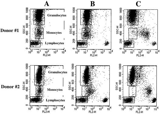
FIG. 1.
Immunofluorescence analysis of the reactivities of CD4 MAbs MT4 and Leu3a with peripheral blood leukocytes. Whole-blood samples were stained with either irrelevant negative control MAb (A), CD4 MAb MT4 (B), or CD4 MAb Leu3a (C) purchased from Becton Dickinson and analyzed by lysed whole blood indirect immunofluorescence. Granularity (SSC) and PE fluorescence (FL2) were plotted to show the binding of the MAb to each leukocyte population. The fluorescence intensities of negative control MAb for all cell populations are marked by rectangles. Two subjects (donor #1 and donor #2) are shown as representative of four studied subjects.

