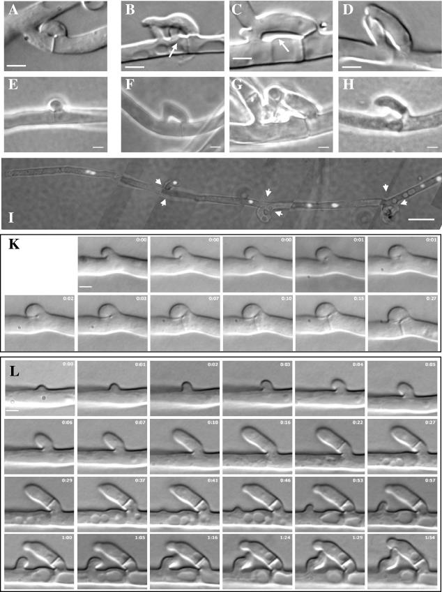
FIG.5.
Clamp connections, clamp-like structures, and pegs in wild-type and Δgap1/Δgap1 dikaryons and nuclear distribution in Δgap1/Δgap1 hyphae. Whereas in the wild type (A) the hook cell fused directly behind the newly synthesized septum, the distances between the septum and fusion site varied in mutant Δgap1/Δgap1 dikaryons (B to D). Arrows mark a peg or weakened cell wall. Pegs could be observed during clamp formation in wild-type (E) and Δgap1/Δgap1 (F to H) dikaryons. The dikaryotic character of Δgap1/Δgap1 hyphae was maintained despite the abnormal mode of clamp-like structure formation (I). The image is composed of seven single images, each representing an overlay of a differential interference contrast and a fluorescence image. Nuclei were stained with DAPI. Arrow heads indicate septae. (K) Time-lapse micrographs of dikaryons during clamp formation in the wild type show the growth of the hook cell continuously directed toward the mother cell (0 to 7 min). The ring visible at the fusion site between the hook and the mother cell (1 to 15 min) indicates that a peg is formed from the mother cell. The ring disappears when the hook and mother cell fuse (27 min). (L) In the Δgap1/Δgap1 dikaryon, the whole process is retarded. The hyphal extrusion representing the future hook initially also grows in the direction of the mother cell (0 to 4 min). Then, it changes its growth direction and grows away from the mother cell (5 to 16 min) and changes its growth direction again a second time (visible at 37 min). A swelling of the cell wall of the mother cell beside the hook indicates the localized weakening of the cell wall at the site of peg formation, where fusion originally was supposed to occur (27 min to 1 h 54 min). Close to this site, a branch develops from the mother cell (41 min) and grows toward the hook cell (41 min to 1 h 54 min) to rescue the failed clamp connection formation. The time is given in hours and minutes. Bars: 2 μm for images A to H, K, and L; 10 μm for image I.

