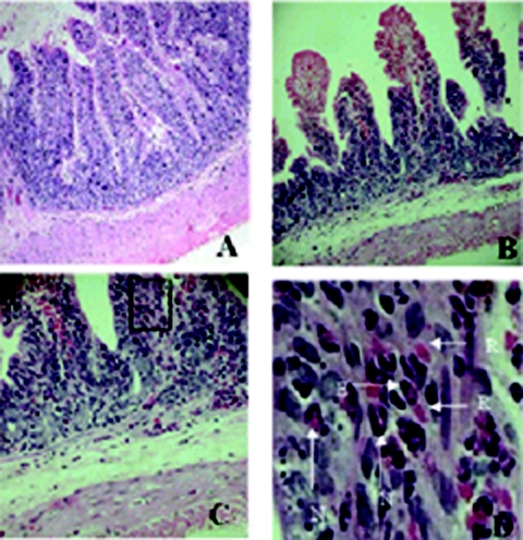
FIG. 5.
Effect of purified HAP in the RIL assay. Purified HAP-treated ileal tissues were processed for histopathological analysis, and photomicrographs were taken. (A) Villous architecture observed in ileal tissues treated with 25 mM Tris-HCl. This photomicrograph shows normal villous structure. Magnification, ×10. (B) Rabbit ileal tissues treated with 40 μg of purified 35-kDa HAP show disruption of normal villous architecture with shortening of the villi. Magnification, ×20. (C) Magnified view of Fig. 4B shows infiltration of polymorphonuclear neutrophils, eosinophils, and erythrocytes. Magnification, ×40. (D) The black box in Fig. 4C was further magnified to show the presence of red blood cells (R), neutrophils (N), and eosinophils (E). Magnification, ×200.

