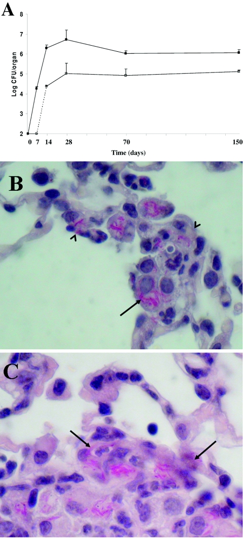
FIG. 3.
Colonization of host tissues following aerogenic challenge with M. tuberculosis strain Erdman (≈100 CFU/mice). (A) Time course of colonization of lung (filled squares) and spleen (empty squares) tissues as assessed by CFU counts. (B and C) Histopathological analysis of lung tissues isolated from infected mice at 14 days postinfection. The left lobe of the lung was removed, fixed, and stained with hematoxylin-eosin and Ziehl-Nielsen stain. Representative slides are shown. Magnification, ×1,000. Arrows indicate AFBs; arrowheads indicate epithelial cells infected with AFBs.

