
FIG. 1.
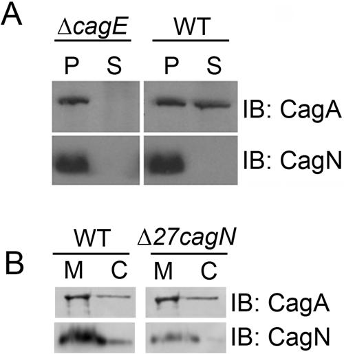
CagN is not delivered into host cells and remains localized to the bacterial membrane. A) Lysates from ΔcagE and wild-type (WT) H. pylori-infected AGS cells were fractionated into a supernatant containing AGS cell cytosol (lanes S) or a pellet containing bacteria (lanes P). B) Fractionated H. pylori lysate shows CagN and Δ27CagN localized mainly at the bacterial membrane (lanes M) rather than the bacterial cytosol (lanes C) as is seen for the inner bacterial membrane-localized protein CagA. IB, immunoblotting.