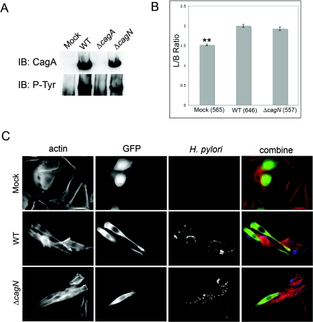
FIG. 3.
A) Western blot showing CagA tyrosine phosphorylation after delivery into host cells. CagA delivery is not disrupted in a cagN mutant. WT, wild type; IB, immunoblotting. B) Image analysis of infected AGS cells shows that wild-type and ΔcagN H. pylori cause similar levels of cell elongation compared to mock-infected control cells. **, significantly different relative to the wild-type H. pylori-infected cells (P < 0.001). Error bars represent standard errors and the numbers of cells counted per condition are in parentheses. L/B, length to breadth. C) Images (magnification, ×600) of infected AGS cells, showing actin, green fluorescent protein-transfected cells, and H. pylori.

