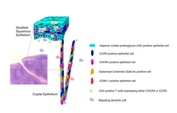
Figure 5.
Schematic representation of cell surface macromolecules and migrating cells implicated in HIV binding and uptake. The inset to the left shows a low magnification photomicrograph of a thin section cut through the external surface of human palatine tonsil (H&E; original magnification: ×40). Most of the external surface of the tonsil is protected by stratified squamous epithelium but there is an abrupt transition to reticulated epithelium at the entrance to a crypt. Cell surface molecules that may contribute to HIV virion binding and the cell types expressing these target molecules are depicted in the diagrammatic representations of stratified squamous epithelium and reticulated cryptal epithelium.

