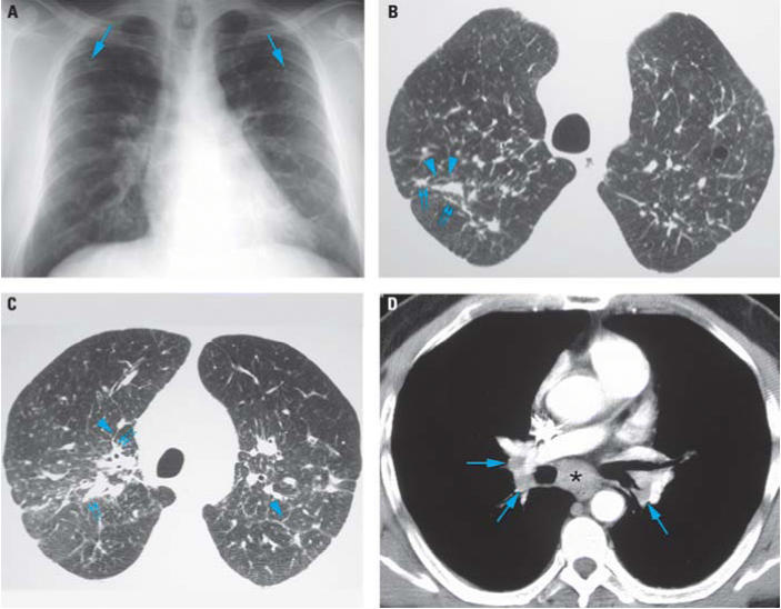
Figure 1.
Chest radiograph (A) and HRCT images (B–D) of patient’s lungs. (A) Frontal chest radiograph showing mild symmetric linear and reticular opacities (arrows) in the upper lobes bilaterally; these opacities are associated with bilateral hilar prominence, suggesting lymphadenopathy. Note the upper lobe distribution of the findings as well as the absence of associated pleural thickening. (B) Axial HRCT image through the lung apices shows bilateral, patchy bronchovascular thickening with a nodular appearance (small double arrows). Nodular interlobular septal thickening is also present (arrowheads). (C) Axial HRCT image through the upper lungs slightly caudal to (B) shows bilateral, patchy bronchovascular thickening with a nodular appearance (small double arrows). Mild interlobular septal thickening is again present (arrowheads). Note the posterior distribution of abnormalities. D) Contrast-enhanced axial CT image shows subcarinal (*) and mild bilateral hilar (arrows) lymphadenopathy.

