
Fig. 1.
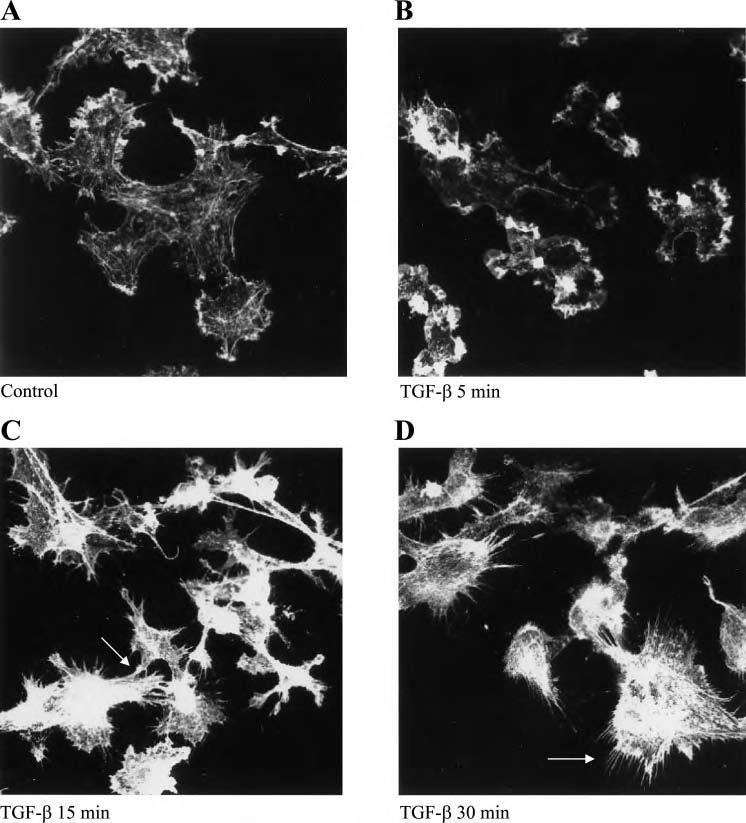
Induction of filipodia formation by transforming growth factor (TGF)-β in human umbilical vein endothelial cells (HUVEC). HUVEC were treated with vehicle or TGF-β1 (10 ng/ml) for various time points and cells were fixed and stained with rhodamine-phalloidin (A-D). Confocal microscopy was used to visualize cells and representative photographs were obtained. There is a dramatic increase in F-actin following TGF-β treatment at 15 and 30 min. Filipodia formation was noted in between cells as well (C) as away from neighboring cells (arrows; D).