
Fig. 5.
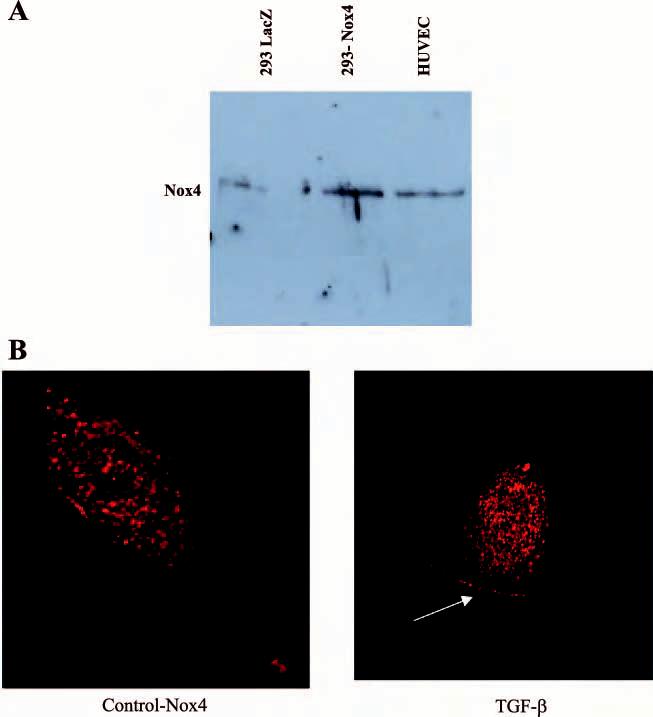
Nox 4 protein in HUVEC. A: cell lysates from 293 cells and HUVEC cells were analyzed for Nox4 protein by Western blot analysis. There is a distinct band at ∼65 kDa corresponding to Nox4 in HUVEC. A faint band is seen in lacZ-transfected 293 cells and is increased with transfection with wild-type Nox4. B: Nox4 protein was visualized by immunostaining of HUVEC with antibody to Nox4 and confocal microscopy. A Bio-Rad MRC-600 confocal laser-scanning microscope mounted on a Zeiss Axiovert 100 fluorescent microscope equipped with a ×63 objective with rhodamine filter was used. Under control conditions, Nox4 is present throughout the cell but concentrated in the nuclear region. With short-term TGF-β treatment, there is increased Nox4 in nuclear compartment and the periphery of the cell (see arrow).