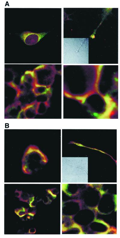
Figure 6.
Colocalization of ankyrin, IP3R-3, and Sig-1R in NG-108 cells. (A) Immunocytohistochemical colocalization (yellow) of Sig-1R (green) and ankyrin B (red) in reticular perinuclear areas (Top Left), plasmalemmal regions of cell–cell contact (lower panels in low and high magnifications, respectively), and growth cones of processes in NG-108 cells (Top Right; Inset: Nomarski optical image). (B) Immunocytohistochemical colocalization (yellow) of Sig-1R (green) and IP3R-3 (red) in perinuclear areas (Top Left), plasmalemmal regions of cell–cell contact (lower panels in low and high magnifications, respectively), and growth cones of processes in NG-108 cells (Top Right; Inset: Nomarski optical image).

