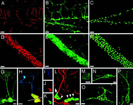
Fig. 1.
Defining neuronal differentiation cascade in the DG. (A) Expression of endogenous nestin, detected by using a monoclonal antibody, in the DG of nestin-GFP transgenic mice; the pattern was the same in WT animals. (B) Expression of GFP in the DG of nestin-GFP transgenic mice; note that endogenous nestin in A is seen mostly in the processes, whereas GFP is present in the processes, the cytoplasm, and the nucleus. Tight packing of cells in the SGZ prevents accurate enumeration of nestin-GFP expressing cells. (C) Expression of CFPnuc, detected by using a polyclonal antibody in the SGZ of nestin-CFPnuc mice; transgene-expressing cells now are represented by their nuclei, thus making accurate cell counts even in densely packed areas possible. (D–F) Expression of nestin (D) and GFP (E) in the SVZ of nestin-GFP mice and CFPnuc (F) in the SVZ of nestin-CFPnuc mice. Note that densely packed SVZ cells, which cannot be accurately counted in D or E, can be quantified easily in F. (G) GFP-expressing neural progenitor cells in the DG of the nestin-GFP mice. The soma of both QNP and ANP cells is seen in the SGZ; QNP cells carry vertical processes, which cross the granule cells layer and end as elaborated arbors in the molecular layer (processes also can be visualized by an antibody to GFAP). ANP cells lack the processes; during and immediately after division, they can be seen in close contact with QNP (note a QNP and an ANP cell above and beneath the dashed line). (H–K) Asymmetric division of stem-like QNP cells in the DG generates ANP cells. (H) After BrdU labeling, cells with GFAP-labeled processes can be seen dividing (note the horizontal plane of division, dashed line) and generating daughter cells that are deposited below, do not carry processes or arbors, and do not stain for GFAP. (I–K) Staining for GFAP (blue; I), BrdU (red; J), and CFPnuc (green; K). (L) A QNP cell (arrow) generates an ANP cell (arrowhead) through an asymmetric division, with the plane of division parallel to the SGZ. Nearby is a cluster of ANP cells (arrowheads) generated through symmetric divisions in the plane perpendicular to the SGZ. GFAP is red, and CFPnuc is green. (M and N) ANPs differentiate into NB1 cells, which start to express markers of young neurons. NB1 cells still are located in the SGZ, cease to express nestin or nestin-CFPnuc, and start to express PSA-NCAM (green), Dcx, and Prox-1. A small subclass of these cells (M) resembles the ANP cells morphologically and is the last cell population to incorporate BrdU. The majority of the NB1 cells (N) extends horizontal processes and does not incorporate BrdU. (O) NB1 cells evolve morphologically and are converted into NB2 cells. NB2 cells are largely confined to the SGZ and extend most of their processes horizontally; however, one process grows vertically or obliquely and extends into the GCL. These cells express PSA-NCAM (green), Dcx, Prox-1, and NeuN. (P) NB2 cells progress into IN. The morphology of these cells resembles that of mature granule neurons. They extend a single vertical apical process and have their soma in the granule cell layer. They express NeuN and Prox-1 but still express PSA-NCAM (green) and Dcx. (Scale bars: A–F, 20 μm; G, H, L–P, 5 μm.)

