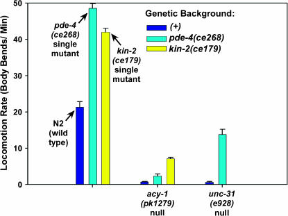
Figure 6.
Loss of PDE-4 partially rescues the paralysis of animals lacking a functional neuronal Gαs pathway. Shown are the mean locomotion rates, expressed as body bends per minute, of strains carrying acy-1(pk1279) (acy-1 null in muscle and nervous system) or unc-31(e928) (unc-31 null). Dark blue bars represent the mutants in a pde-4(+) (wild type for pde-4) background, cyan bars in the second and third sets represent double mutants carrying the indicated mutations in the pde-4(ce268) (strong reduction-of-function) background, and the yellow bar in the second set represents the acy-1(pk1279); kin-2(ce179) double mutant. Wild-type animals and single-mutant controls are shown in the first set of three bars. Error bars represent the standard error of the mean for 10 animals. Values for acy-1(pk1279) and kin-2(ce179) are reprinted with permission from Schade et al. (2005); values for unc-31(e928) single mutants are reprinted with permission from Charlie et al. (2006).

