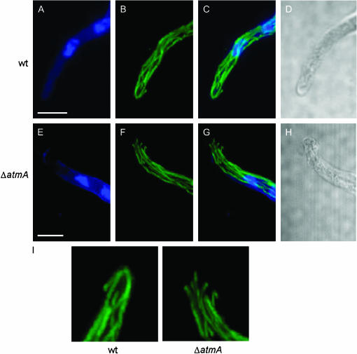
Figure 8.
Microtubules fail to converge at the hyphal tip in ΔatmA mutants. Wild-type (A–D) and ΔatmA (E–H) hyphae were examined by immunofluorescence microscopy. (A and E) Nuclei visualized using Hoechst 33258. (B and F) Microtubules visualized with an anti-β-tubulin antibody. (C and G) merged image. (D and H) Brightfield images (single section, whereas all other images are maximum projection of Z stacks). (I) The microtubule organization at the hyphal tip is enlarged to highlight the defect. Bars, 5 μm.

