Figure 3.
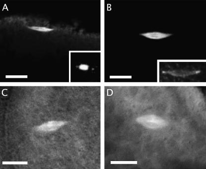
Meiosis I in stage 14 oocytes. (A and B) Fixed oocytes stained for α-tubulin. Both mud4 (A) and wild-type (B) oocytes display a bipolar anastral spindle with focused poles that is oriented parallel to the oocyte surface. Staining of a mutant ooctye for DNA (inset in A; not the same oocyte as in A) reveals that chromosome positioning is normal, with the nonexchange fourth chromosomes visible away from the center. Staining of a wild-type oocyte for Mud (inset in B; the same oocyte as in B) shows that the protein is localized at the poles. (C and D) Unfixed oocytes from lines bearing an α-tubulin–GFP transgene examined by confocal microsopy. The spindle of both mud4 (C) and wild-type (D) oocytes is of normal shape and position. Bars, 10 μm.

