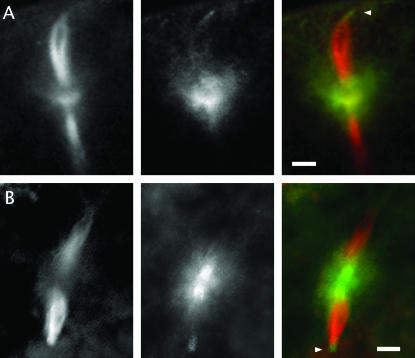
Figure 6.
The distribution of Mud protein in wild-type eggs undergoing meiosis. Freshly laid eggs (whose cortex is oriented toward the top of the page) stained for α-tubulin (left column; red in composite) and Mud (middle column; green in composite) are shown. In these respresentative examples of eggs in meiosis II, Mud protein is found in a broad belt that includes and surrounds the central pole body. It is also expressed at the anastral poles (marked by arrowheads when in focus) of the meiosis II apparatus. (A) An example with the same anti-Mud antibody as used in Figure 5. (B) An example with an anti-Mud antibody directed against a more amino-terminal portion of the protein (see Figure 1). Bars, 10 μm.

