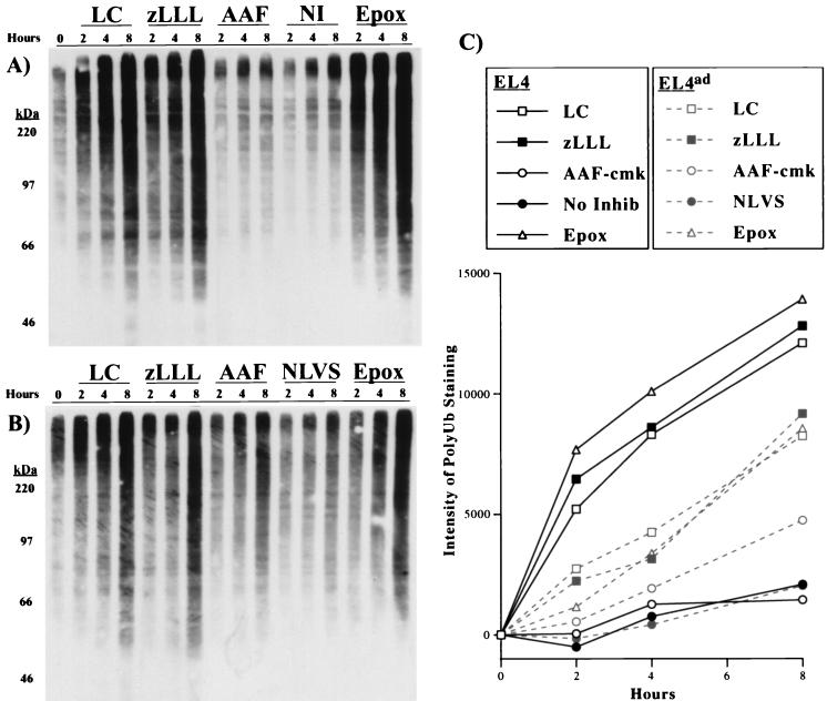
Figure 2.
Effect of proteasome inhibitors on the accumulation of polyUb proteins in EL4 and EL4ad cells. (A and B) EL4 (A) and EL4ad (B) cells treated with inhibitors for 2, 4, and 8 h were analyzed by Western blotting with the use of the FK2 mAb, the binding of which was visualized by chemiluminescence. The inhibitors used were 10 μM lactacystin (LC), 10 μM zLLL (zLLL), 10 μM AAF-cmk (AAF), no inhibitor (NI), 50 μM NLVS (NLVS), and 1 μM epoxomicin (Epox). (C) The increase in FK2 staining was quantitated and is represented graphically.

