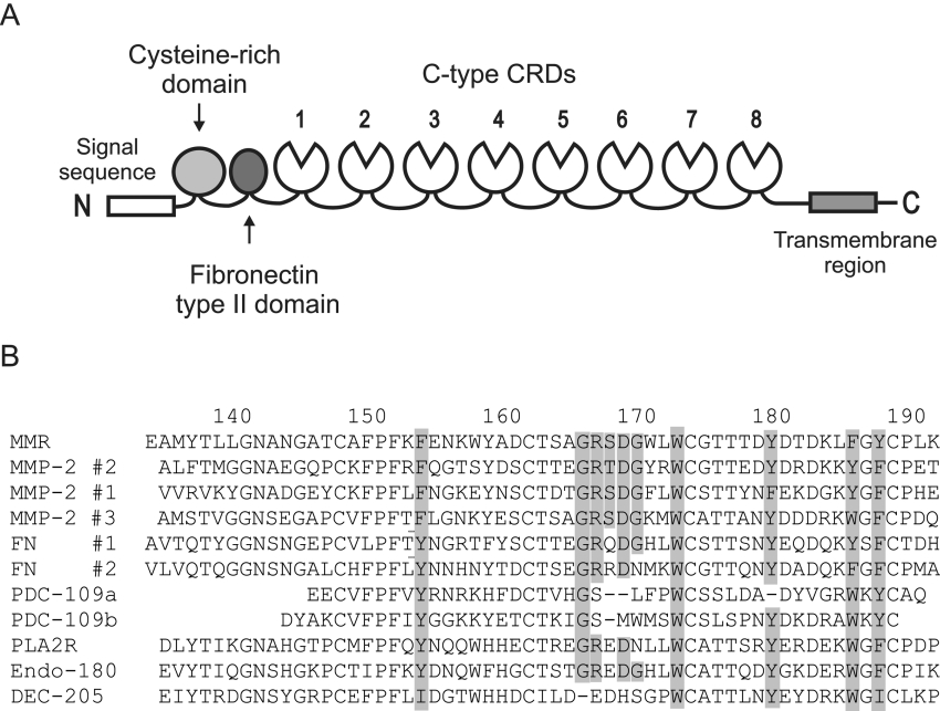
Figure 1. Domain organization of the mannose receptor.
(A) Diagrammatic representation of the mannose receptor. (B) Sequence alignment of selected FNII domains. MMP-2 #1, #2 and #3, three domains from MMP-2; FN #1 and #2, first and second domains from fibronectin; PDC-109a and b, two domains from bovine seminal plasma protein PDC109; PLA2R, phospholipase A2 receptor. Residues predicted to form the collagen-binding region in MMP-2 #2 [23,24] are shaded in MMP-2 #2 and where they are conserved in other domains. Numbers at the top correspond to the residues of the mannose receptor.

