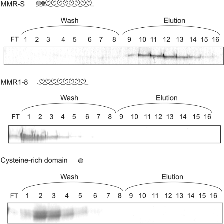
Figure 2. Binding of mannose receptor fragments to gelatin.
Mannose receptor fragments were loaded on to a gelatin–agarose column (2 ml). Wash and elution fractions were analysed by SDS/PAGE (15% gels for MMR-S and MMR1–8, and 17.5% for the cysteine-rich domain). Gels were stained with Coomassie Blue. FT, flowthrough.

