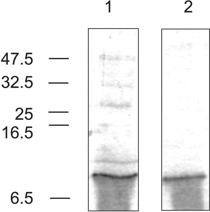
Figure 3. Expression and purification of mannose receptor FNII domain.

Expressed FNII domain purified by Ni-NTA–agarose affinity chromatography (lane 1) and gelatin–agarose affinity chromatography (lane 2). Gels (20% polyacrylamide) were stained with Coomassie Blue. The positions of molecular-mass markers are indicated on the left (sizes in kDa). The smearing seen under the band is a gel artefact that is often seen when running very small proteins using the Tris/glycine buffer system. N-terminal sequencing of material taken from the smear indicates that it is intact FNII domain.
