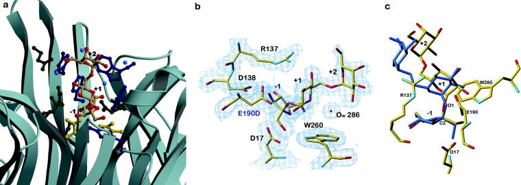
Figure 3. Localization and structural arrangement of raffinose bound in the active-site pocket in Tm invertase.
(a) Ribbon representation of the active-site pocket in Tm invertase. The raffinose molecule trapped in the active site is shown in coloured atoms as a stick-representation. (Oxygen atoms are coloured in red). Catalytic residues are shown in gold, aromatic residues in dark blue and other residues in green. Blue balls are water molecules. The substrate-binding subsites are annotated from −1 to +2. (b) Electron density calculated after the final refinement step for the Tm invertase inv-E190D in complex. The Fourier map (2Fo–Fc) is contoured at 1σ showing the trapped substrate molecule present in the active site of inv-E190D. (c) Stick representation of raffinose in complex with Tm inv-E190D (standard atom type colours) superimposed on to the crystal structure of raffinose pentahydrate (in blue). Figures 3(b) and 3(c) were prepared with TURBO-FRODO [19].

