
Figure 3. RH-123 fluorescence changes in digitonin-permeabilized lymphocytes upon addition of respiratory substrates and inhibitors.
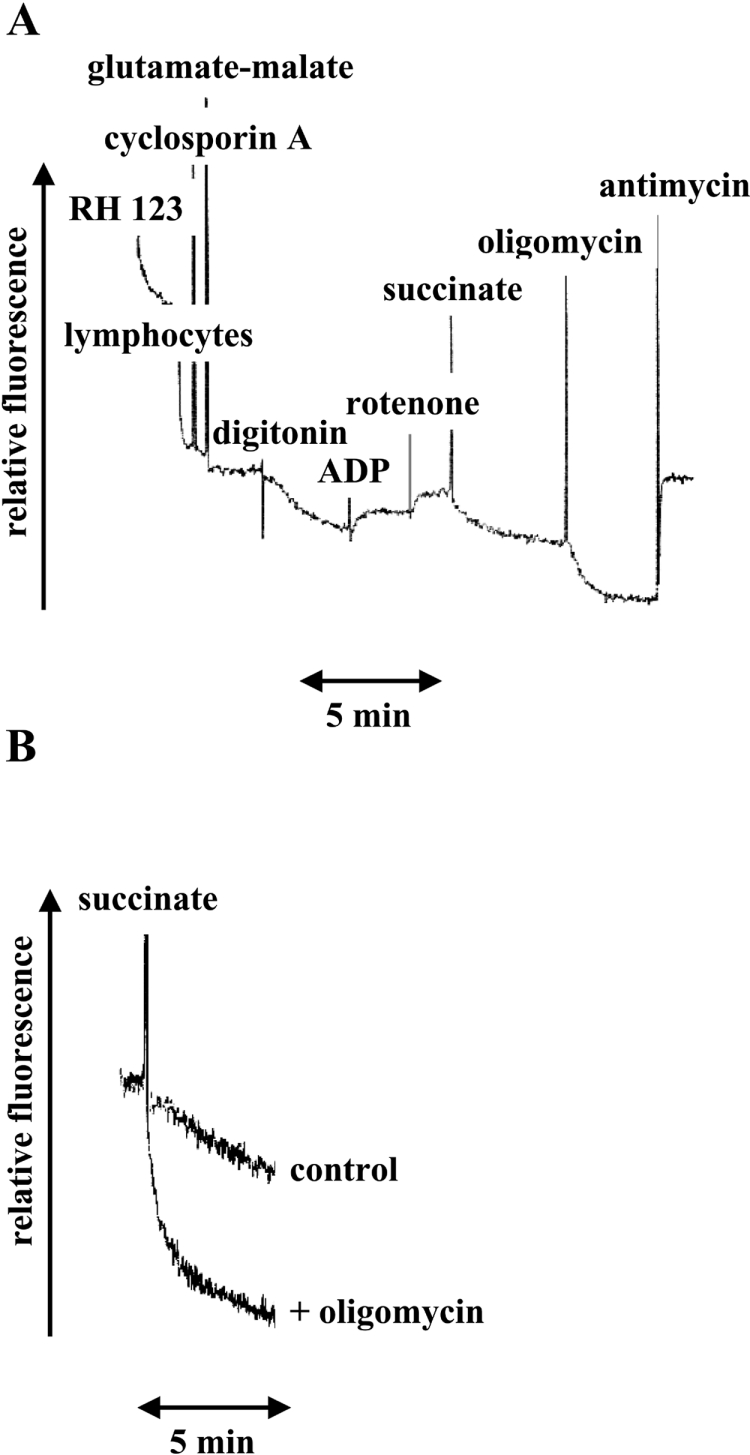
(A) Fluorescence measurements were carried out in a respiratory buffer (250 mM sucrose, 10 mM Hepes, 100 μM K-EGTA, 2 mM MgCl2 and 4 mM KH2PO4 (pH 7.4), containing an ADP regenerating system (10 mM glucose and 2.5 units of hexokinase). Additions were 50 nM RH-123, 2×10⁶/ml lymphocytes, 33 nM cyclosporin, 10 mM glutamate/10 mM malate, 15 μg/ml digitonin, 200 μM ADP, 2.5 μM rotenone, 20 mM succinate, 2 μM oligomycin and 1 μg/ml(1.8 μM) antimycin. (B) RH-123 fluorescence kinetics induced by the addition of 20 mM succinate to digitonin-treated lymphocytes pre-incubated or not with 2 μM oligomycin. The sample mixture consisted of 0.5 ml of respiratory buffer containing an ADP regenerating system, 50 nM RH-123, 2×10⁶/ml lymphocytes, 33 nM cyclosporin, 2.5 μM rotenone, 0.2 mM ADP and 15 μg/ml digitonin.