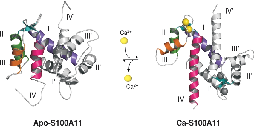
Figure 2. Calcium-dependent conformational change in S100 proteins.
The three-dimensional structures of calcium-free S100A11 (apo-S100A11) and calcium-bound S100A11 (Ca-S100A11) are shown to demonstrate the calcium-induced conformational change. In the symmetrical dimer, helices of one monomer (I–IV) are highlighted in different colours, while the other monomer (helices I'–IV') is coloured grey. As sensors, the S100 proteins experience a conformational change upon calcium binding (four atoms/dimer). The rearrangement of helix III (orange) exposes previously buried residues that are essential for target recognition (not shown, see Figure 3) and further biological response. This Figure was drawn using MacPyMOL (http://delsci.com/macpymol/).

