
Fig. 1.
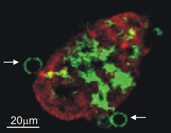
Lamina propria DCs extend trans-epithelial processes with a “balloon-body” appearance. The terminal ileum of a CD11c-EGFP mouse was isolated and the lumen exposed. Villous epithelial cells were stained using the red fluorescent dye SNARF and the tissue imaged by two-photon microscopy. A reconstructed transverse section shows a single z slice from one villus with 2 “balloon bodies” formed by DC trans-epithelial membrane extensions marked by white arrows. The cropped field is 170 μm × 133 μm from an original field of 560 μm × 560 μm.