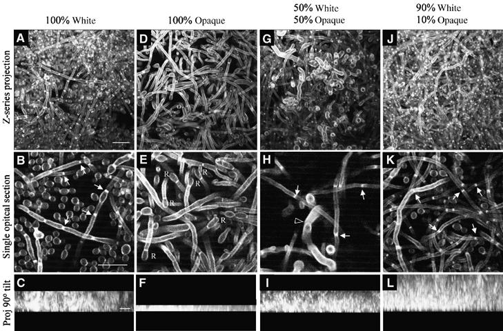
Figure 7.

A minority of opaque cells enhance white cell biofilm development. Different proportions of white and opaque cells were mixed and the same total number of cells spread on a silicon elastomer. After 90 min of undisturbed incubation, the elastomers were gently rinsed, then flooded with fresh growth medium and gently rocked for 48 h. In all combinations, white cells were 50% a/a and 50% α/α, and opaque cells 50% a/a and 50% α/α. In all combinations, a monolayer of cells covered the elastomer at the initiation of rocking which reflected the proportions of white and opaque cells initially inoculated. (A, D, G, J) Z-series projection of multiphoton LSCM scans through the biofilm. (B, E, H, K) Single optical section in the middle of the biofilm. (C, F, I, L) Z-series projections viewed from the side (90° tilt). At the end of the incubation period, the top of each gel was identified by a precipitous decrease in pixel intensity. The small solid arrows in (B), (H) and (K) point to septae in hyphae. The unfilled arrowhead in (H) points to a conjugation tube. ‘R's in (E) refer to apical reversion to the budding growth form at the ends of conjugation tubes that have failed to fuse. Scale bar in the first image for each horizontal row represents 10 μm.
