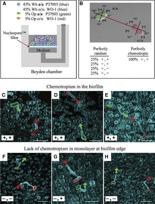
Figure 8.

Majority white cell biofilms promote chemotropism between rare opaque a/a and opaque α/α cells. (A) Diagram of the modified Boyden chamber that was employed. The upper well was inoculated with 45% white a/a cells, 45% white α/α cells, 5% opaque a/a cells vitally stained green with fluorescein-conjugated ConA and 5% opaque α/α cells vitally stained red with rhodamine-conjugated ConA. Medium was replenished from below after 24 h. After 48 h at 29°C, cells were fixed, stained with calcofluor (Blue) and imaged by multiphoton LSCM. Cells were visualized in the upper region of the thickest portion of the biofilm or in the monolayer at the biofilm edge. Fields were scanned for a green and red cell that had formed conjugation tubes within 25 cell diameters of each other. (B) The orientation of the conjugation tubes was assessed as diagrammed. (C–E). In the majority of cases in which a red and green cell were observed within 25 cell diameters of one another (18 of 20; 90%) in the three-dimensional upper region of the biofilm proper, their conjugation tubes were oriented in the approximate direction of each other (+, +). (F–H) In the majority of cases in which a red and green cell were observed within 25 cell diameters of one another (16 of 20; 80%) in the two-dimensional monolayer at the edge of the biofilm proper, one or both tubes were oriented away from each other (+, − or −, + or −, −). a/a cells, strain P37005; α/α cells, strain WO-1. Scale bar, 5 μm.
