
Figure 1.
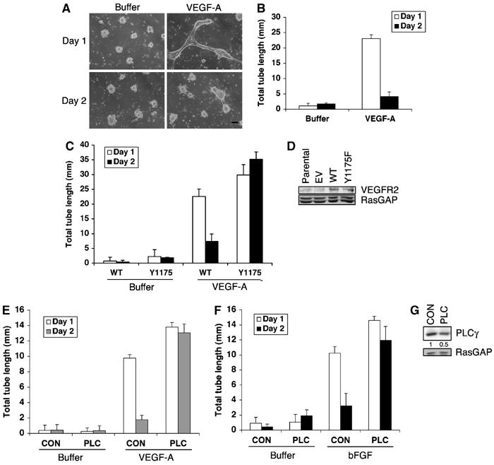
(A, B) Tubes formed in the presence of VEGF-A, and then spontaneously regressed. BRECs were plated in a collagen sandwich gel, and then medium containing 2.5 ng/ml VEGF-A or buffer was added. The medium was changed every day. At the indicated times, three randomly selected fields were photographed and tube lengths were measured (B). Representative photos are shown. Bar, 100 μm (A). The bar graph is the mean±s.d. of 3 wells/treatment. (C, D) Tubes failed to regress when the cells expressed a VEGFR2 mutant that was unable to activate PLCγ. BRECs expressing WT or mutant VEGFR2 (Y1175F) were subjected to a tube assay. A Western blot of total cell lysates shows that the expression level of the introduced WT and Y1175F VEGFR2 was similar (D). (E–G) Reducing the level of PLCγ-stabilized tubes. HUVECs were transfected with either siRNA for PLCγ (PLC) or non-targeting control siRNA (CON) and subjected to a tube assay in the presence of 2.5 ng/ml VEGF-A (E) or 25 ng/ml bFGF (F). Total cell lysates were subjected to Western blot analysis using an anti-PLCγ antiserum. The blot was reprobed with an anti-RasGAP antiserum to verify equivalent protein loading. PLCγ expression was decreased by an average of 50% in siRNA PLCγ-transfected cells (G).