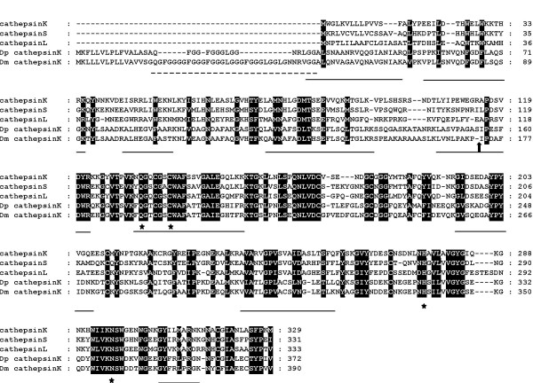
Figure 2.
ClustalX amino acid sequence alignment of D. melanogaster cathepsin K (Dm cathepsinK, CG4847), D. pseudoobscura cathepsin K (Dp cathepsinK, GA18475) and human cathepsin K, S, and L. Active site residues, stars; Gly-rich region, dashed line; predicted cleavage to generate mature cathepsin, filled block arrow; tryptic peptides identified by mass spectrometry, underline. Conserved amino acids are in black.

