
Figure 1. Signal-Transduction Pathways Involved in Resistance of C. trachomatis-Infected Cells against Apoptosis.
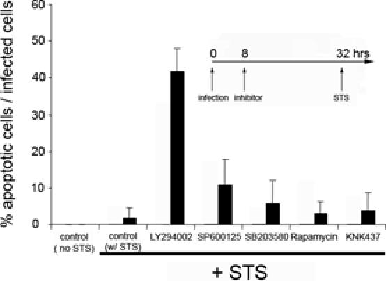
All cells were infected with C. trachomatis at an MOI of 1.0 for 32 h and then treated overnight with 2 μM STS. In uninfected cells that had been treated with STS under the same conditions, 83 ± 10% of the cells had apoptotic nuclei. Inhibitors were added 24 h before STS treatment, as indicated in the inset. The inhibitor concentrations (and targeted mediators) were: 50 μM LY294002 (PI3K), 10 μM SB203580 (P38), 10 μM U0126 (MEK5), 10 μM SP600125 (JNK1/2), 5 nM rapamycin (mTOR), and 50 μM KNK437 (heat shock proteins). The “control (no STS)” contained infected cells, while “control w/STS” contained infected cells that were incubated with buffer without inhibitor and then treated with STS. DNA was revealed by Hoechst staining, and values are expressed as the % of infected cells that had apoptotic nuclei. The average and SD were calculated from the values obtained from at least 50 microscope fields. The experiment was performed on three separate days, and results from a representative experiment are shown.