
Figure 8. Localization of 14-3-3β in C. trachomatis-Infected Cells.
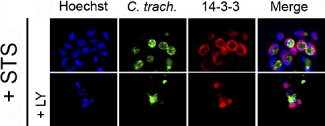
Cells expressing endogenous levels of BAD were infected with C. trachomatis at an MOI of 1.0 for 26 h, and then incubated with 50 μM LY294002 (LY) or control buffer for 6 h before treating with 2 μM STS overnight. DNA was revealed with Hoechst (blue), Chlamydia with FITC-conjugated anti-Chlamydia antibody (green), and 14-3-3 with Texas Red-conjugated anti-14-3-3 antibody (red), as described in Materials and Methods. 14-3-3 is localized primarily on the Chlamydia vacuole in infected cells, even in the presence of STS (top row). The co-localization of 14-3-3 and the Chlamydia vacuole is lost in infected cells that had been pre-incubated with LY294002 before STS treatment (bottom row). One experiment of three representative experiments performed on separate days is shown.