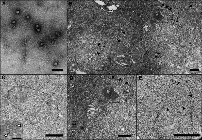
Figure 5. Wolbachia Infecting the Testes of N. vitripennis .
(A) Negatively stained virus particles of uniform size from PEG-precipitated preparations of A-infected N. vitripennis adults. No tail-like structures were apparent, perhaps due to disruption during the purification process. Bar = 100 nm.
(B) Low-resolution transmission electron micrograph of individualized spermatids and Wolbachia in N. vitripennis pupal testes. Ax and Md denote spermatid axonemes and mitochondrial derivatives. Several of the noted Wolbachia cells (W) are shown in higher resolution in Figure 5C–5E. Solid arrowheads denote phage particles. Bar = 200 nm.
(C) A high density of phage particles within Wolbachia is shown; phage tails are occasionally visible and noted by white arrowheads in the inset. Bar = 200 nm; inset bar = 100 nm.
(D) Virion-free (lower right) and virion-containing Wolbachia (upper right) localized near two N. vitripennis spermatids are shown. Solid arrowheads denote phage particles inside Wolbachia, and Ax and Md denote spermatid axonemes and mitochondrial derivatives, respectively. Bar = 200 nm.
(E) Solid arrowheads denote phage particles dispersing from within putatively lysed Wolbachia into the extracellular matrix. Bar = 200 nm.

