
Figure 3.
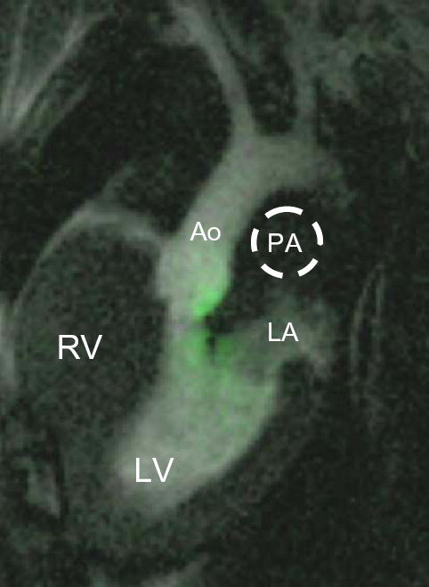
Selective real-time MR angiogram confirming ASP needle entry into the left atrium. After visual confirmation of ASP needle entry in the left atrium, 3–5mL of 30mM dilute Gd-DTPA is injected through the ASP needle wire port. A saturation pre-pulse is applied to suppress the background as contrast sequentially enhances the lumen of the left atrium (LA), left ventricle (LV) and out the aorta (Ao). Note there is no contrast enhancement of the right ventricle (RV) or outlined pulmonary artery (PA), indicating successful ASP.