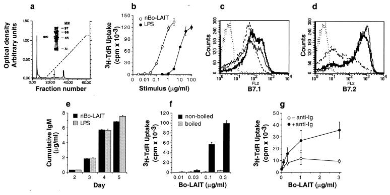
Figure 1.
Bo-LAIT stimulates the growth and differentiation of resting B cells. (a) The chromatograph of the last step of Bo-LAIT purification on hydroxy-apatite. Arrow indicates the peak bioactive fraction. The left track of the Inset depicts SDS/PAGE of 5 μg of protein derived from the peak bioactive fraction followed by silver staining. (b) Comparative B cell growth promoting activity of Bo-LAIT and LPS. (c and d) Bo-LAIT induced up-regulated expression of B7.1 and B7.2. Dotted line, isotype control; dashed line, unstimulated cells; solid line, 50 μg/ml LPS; bold line, 0.3 μg/ml Bo-LAIT. (e) Comparative differentiation promoting activity of 0.5 μg/ml Bo-LAIT and 50 μg/ml LPS. (f) Heat lability of nBo-LAIT. Samples were boiled or not for 10 min and used to stimulate high buoyant density murine splenic B cells. (g) Growth-promoting activity of nBo-LAIT on human tonsil B cells in the presence (+) or absence (−) of a submitogenic concentration of immobilized mAbs specific for human Igκ and Igλ.

