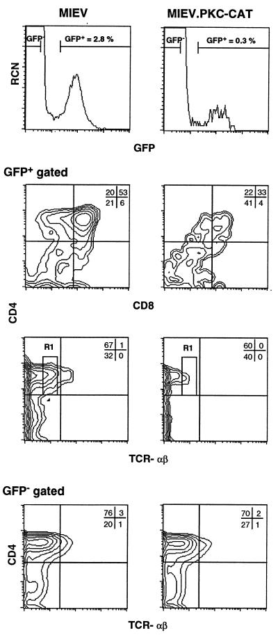
Figure 4.
PKC-derived signals arrest T cell development at the DP stage. Intact fetal thymic lobes from d14 timed-pregnant CD1 mice were retrovirally infected with either vector alone (MIEV) or PKC-CAT encoding constructs for 72 h in HOS-FTOC, then incubated in standard FTOC for 5 days and analyzed for GFP expression (Top). Flow cytometric analyses of CD4 vs. CD8 and CD4 vs. TCR-αβ cell surface expression on infected (GFP+-gated) cells (Middle) and (GFP−-gated) cells (Bottom) are shown. The frequency of GFP+ thymocytes with a TCRlo phenotype, R1-gate, from PKC-CAT- and MIEV-infected FTOCs is 6% and 10%, respectively. The data shown are representative of three independent experiments.

