Figure 2.
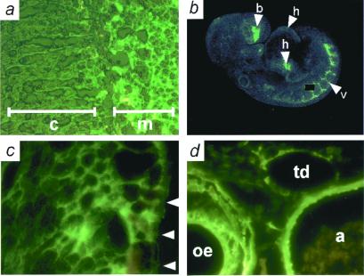
Sites of Adm expression determined by EGFP fluorescence. (a) Frozen section of adrenal gland from an adult Adm+/−. Note intense fluorescence in adrenal medulla (m) and the absence of fluorescence in adrenal cortex (c). (b) Confocal microscopy of an E9.5 Adm+/− embryo. Moderate expression is seen in the heart (arrowhead h), and strong expression in the developing vasculature (arrowhead v) and forebrain (arrowhead b); a speck of nonspecific fluorescence has been covered with a black box. (c) Frozen section of an E13.5 Adm+/− embryo, showing expression in the developing left ventricle. Arrowheads indicate the pericardial surface of the left ventricle. (d) Frozen section of an E16.5 Adm+/− embryo, showing expression in the aorta (a), esophagus (oe), and thoracic duct (td).

