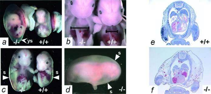
Figure 3.
Adm−/− embryos have massive generalized edema. (a) Appearance of E14.5 Adm−/− (Left) and Adm+/+ (Right) embryos in their yolk sacs shortly after dissection from the uterus. Note that the yolk sac (arrowhead ys) of the Adm−/− embryo is distended with fluid, and its blood vessels are thinner than those of its wild-type littermate. (b) The thoracic cavity of E13.5 Adm−/− embryos is considerably enlarged (Left) compared with a wild-type littermate (Right). The black lines indicate the width of the thoracic cavity just above the diaphragm. (c) Severe hydrops fetalis is apparent in the E14.5 Adm−/− embryos (Left), readily visible after their yolk sacs are removed. The arrowheads (s) indicate the outer skin layer of the embryos. (d) The extreme hydrops of the Adm−/− embryos, dissected away from their yolk sacs, is completely general, as indicated by the uniformly swollen back and head. The arrowheads (s) again indicate the outer skin layer. (e and f) H&E stain of transverse sections through E14.5 Adm+/+ (e) and Adm−/− (f) embryos. The Adm−/− embryo has a fluid-filled thoracic cavity, and the tissues external to the rib cage are markedly swollen (e and f, ×1).

