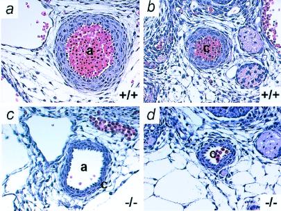
Figure 5.
Adm−/− embryos have thin arterial walls. Transverse sections through vessels of Adm+/+ (Upper) and Adm−/− (Lower) embryos at E13.5 were stained with H&E. (a and c) aorta; (b and d) carotid artery. Note the thin vascular walls (approximately three cells thick) of the Adm−/− vessels compared with wild-type vessels (approximately five cells thick). a, aorta; c, carotid artery (×10).

