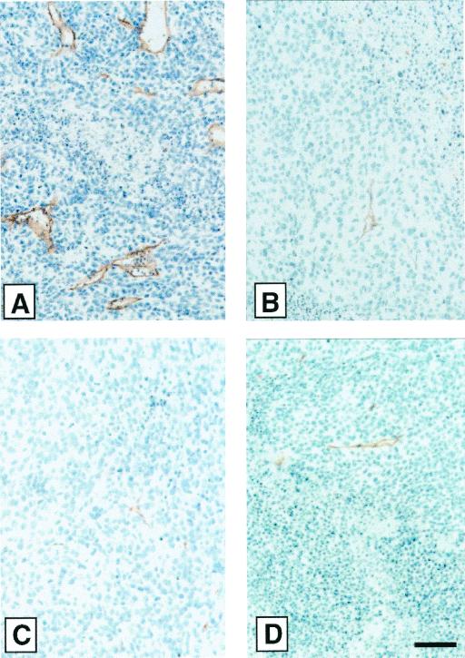
Figure 5.
Immunohistochemical staining for blood vessels in sections of KRIB tumors. KRIB osteosarcoma tumors from a treatment experiment similar to the one shown in Fig. 3 were removed at the end of the experiment and sectioned, and the sections were stained with anti-CD31 antibodies to visualize tumor blood vessels. Representative microscopic fields from the tumors show a higher density of blood vessels in the vehicle alone group (A) than in the anastellin (B), sFN (C), and sFBG (D) groups (Magnification, ×400; bar = 50 μm.)

