Figure 3.
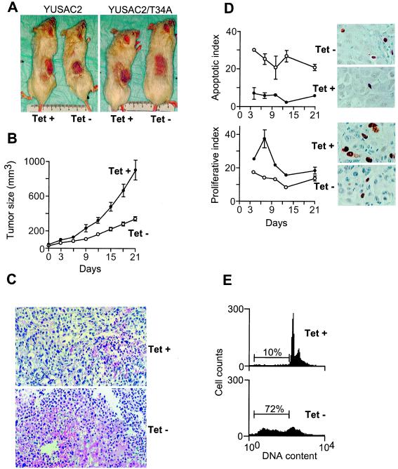
Effect of survivin Thr34→Ala mutant in melanoma tumors in vivo and tet-regulated apoptosis of tumor lines reestablished in vitro. (A) Untransfected YUSAC2 cells (Left) or YUSAC2/T34A-C4 cells (Right) were injected s.c. into CB.17 mice, and tet was added (left side of images) or withheld (right side of images) from the drinking water as indicated. Photographs were taken of representative mice 8 weeks after injection. (B) YUSAC2/T34A-C4 tumors were established in 30 animals maintained on tet. When tumors became apparent (day 0), tet was maintained in the drinking water of 10 animals (tet+, ●) and withheld from 20 animals (tet−, ○), and tumors were monitored for 3 weeks. Bars = SD, P < 0.0001 for days 12–21 by two-tailed t test. (C) Histology of established YUSAC2/T34A-C4 tumors from tet+ (Upper) and tet− (Lower) animals after 8 weeks by hematoxylin/eosin staining (×100). (D) YUSAC2/T34A-C4 tumors were established in animals on tet, and when tumors became apparent (day 0), tet was maintained in the drinking water of half the animals (tet+, ●) and withheld from the others (tet−, ○). Tumors were excised on days 4, 7, 10, 13, and 21 and subjected to TUNEL and BrdUrd staining for respective determination of apoptotic (upper graph) and proliferative (lower graph) indices. Bars = SEM from 2 animals for days 4 and 13, 4 animals for days 7 and 10, and 10 animals for day 21. Adjacent images are representative fields (×400) from day 7 tumors ± tet as indicated. (E) Cell lines from several YUSAC2/T34A-C4 tumors slowly growing in vivo after withdrawal of tet were reestablished in vitro. The DNA content analysis for a representative line after culturing for 72 h in the presence (Upper) or absence (Lower) of tet is shown. The marker and percentages indicate the sub-G1 fraction, corresponding to apoptotic cells.

