
FIG. 5. Structural models of the dynein stalk.
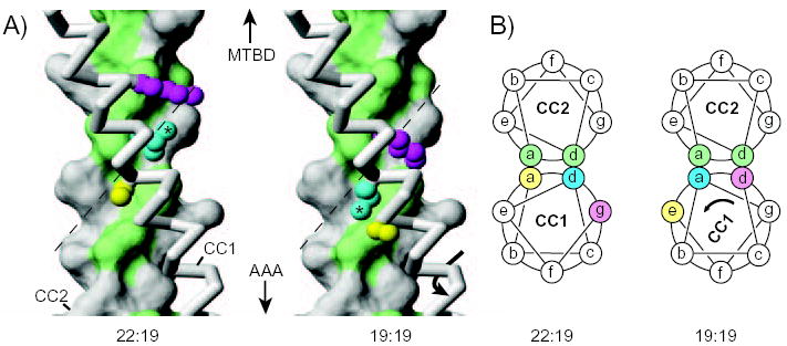
A, Models of a section of the dynein stalk in configurations expected to correspond to those occurring in SRS-22:19 or SRS19:19. CC2 is shown in surface representation with hydrophobic residues (Val, Ile, Leu, Ala, Met, Tyr) in the coiled-coil core (“a” and “d” positions) coloured green. CC1 is shown in backbone representation (grey), with side chains included for Ser3224 (yellow), Leu3227 (cyan; with asterisk) and Lys3230 (magenta). In passing from the configuration of SRS-22:19 to that of SRS-19:19, the side chain of Leu3227 shifts from packing against one side of CC2 (left) to packing against the other side (right), potentially by following the hydrophobic-lined groove, formed by Ile3459, Leu3463, Val 3466 and Val 3470, in the surface of CC2 (dashed line). Similar hydrophobic grooves in the core interface of CC2 occur in most other heptads along the length of the stalk coiled coil (Supplemental Data). B, Cartoons depicting transverse sections through the coiled-coil stalk in the region shown in Fig. 5A. Residues in CC1 are coloured as in Fig. 5A to illustrate that Leu3227 (cyan) shifts from an “a” heptad position to a “d” position in going between the 22:19 and 19:19 configurations (see also Fig. 2A). Part A of this figure was prepared with the program MOLMOL (28).