
Figure 4.
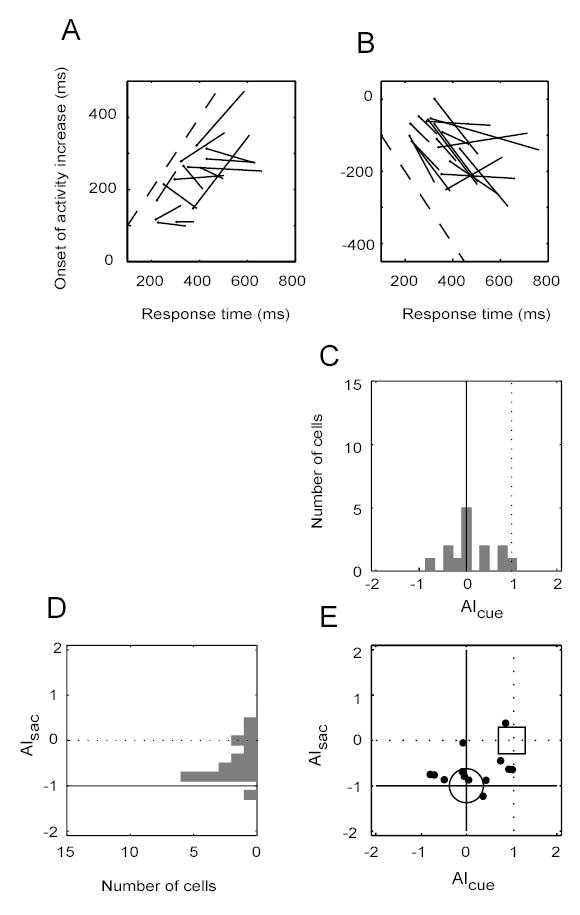
Timing of target discrimination (IN vs. OUT) in cell activity. (A) Data are aligned on cue onset. The response times (RTs) for each cell were divided into three trial groups (shortest, middle and longest RTs). Each line shows the linear regression of median activation times on median RTs for one cell. The filled circle at the end of each line is the end of the regression line for the shortest RT trial group. The dashed line has unity slope. Other details of the plot are the same as in Figure 2A. (B) Data are aligned on saccade onset. Otherwise the plot is the same as in (A). (C) The distribution of the line slopes in (A) is shown in a histogram. AIcue denotes the line slopes in (A). (D) The distribution of the line slopes in (B), or AIsac, is shown in a histogram. (E) An X–Y plot of the joint distributions from (C) and (D). The large circle indicates the expected location of the slopes if the timing of target discrimination in a cell’s activity is time-locked to the cue. The square indicates the location if the discrimination is time-locked to the saccade.