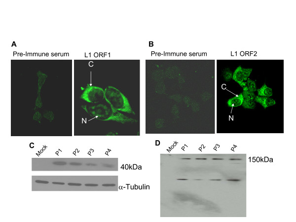
Figure 2.
RC-L1 is expressed in breast cancer cells. RC-L1- expression determined by immunofluorescence using anti-L1 ORF1 (A) and anti-L1 ORF2 (B) rabbit polyclonal antibodies showed cytoplasmic "C" and nucleolar "N" staining. No specific staining was detected when using pre-immune serums as control. Immunoblotting of whole cell lysates using anti-ORF1 (C) or anti-ORF2 (D) detected a 40 kD and a 150 kDa respectively. Additional bands were detected by anti-ORF2 antibody at 135 kDa and 66 kDa which may be due to cleavage by a cellular protease. P1 through P4 correspond to consecutive passages of RC-L1 expressing cells. Anti-α-tubulin was used as a loading control. Mock transfected cells serve as a negative control.

