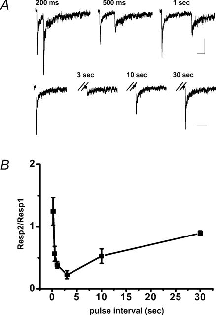
Figure 1. Paired-pulse activation of nAChRs in hippocampal interneurones is potentiated at a 200 ms pulse interval.
A, representative traces of α7-containing nAChR responses from a single CA1 hippocampal interneurone evoked by photolysis of cCarb (500 μm) by 15 ms UV laser pulses at varying intervals. The interpulse intervals are shown above the traces; for the bottom traces, the interval between the first pulse (shown on the left) and second are indicated above the hash marks. Scale bars are 50 pA and 250 ms. B, plot of the ratio of the amplitude of the response evoked by the second uncaging pulse to the first uncaging pulse (Resp2/Resp1) versus pulse interval. Data are means ± s.e.m. from 7–10 interneurones for each data point.

