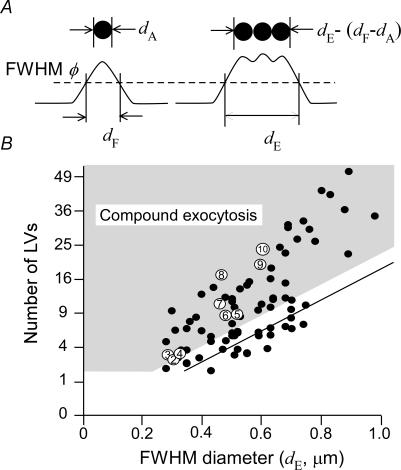
Figure 5. Relationship between numbers of LVs and FWHM diameters in multi-step and massive exocytic events in PC12 cells.
A, relationship between actual diameters of vesicles (upper panels) and their FWHM diameters of fluorescence profiles (lower panels). B, the number of LVs involved in each exocytic event plotted against its FWHM diameter, which was estimated by dividing the volume of massive events by that of single events (0.005 μm3). The straight line denotes the maximal number of LVs that can be explained by surface vesicles [(dE−dF+dA)/dA]2. The shaded area represents the events that cannot be accounted for by surface exocytosis at the plasma membrane, taking into account an error of 0.1 μm in the estimates of FWHM diameters. Open circles with a number denote data obtained from the events shown in Fig. 1B.

