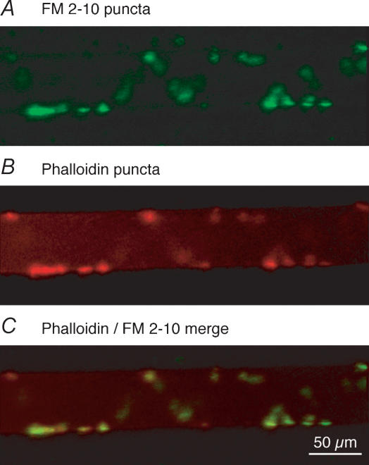
Figure 3. Recycling vesicle clusters colocalize with actin bundles at the presynaptic terminal.
A giant axon was impaled with a microelectrode first containing KCl (1 m), and after FM staining with a second electrode containing labelled phalloidin (Alexa Fluor 568 phalloidin). FM 2-10 was applied to the ventral surface of the spinal cord as a continuous stream from a pipette (see Fig. 1A). A, FM2-10 was loaded into synaptic vesicles in lamprey giant axons by repetitive stimulation of a single axon through the KCl intracellular electrode in the presence of extracellular FM dye. On imaging the tissue, puncta of dye are observed along the periphery of the recorded axon. B, following the FM loading step, the same axon was re-impaled with a phalloidin-containing electrode. Presynaptic injection of fluorescent phalloidin into the same axon reveals location of actin clusters surrounding vesicles. C, overlay of A and B demonstrates that the location and structure of the phalloidin-labelled actin bundles (B) show an identical distribution to the vesicle clusters (A). In a total of 5 preparations, all observable puncta (n = 40) showed colocalization.

