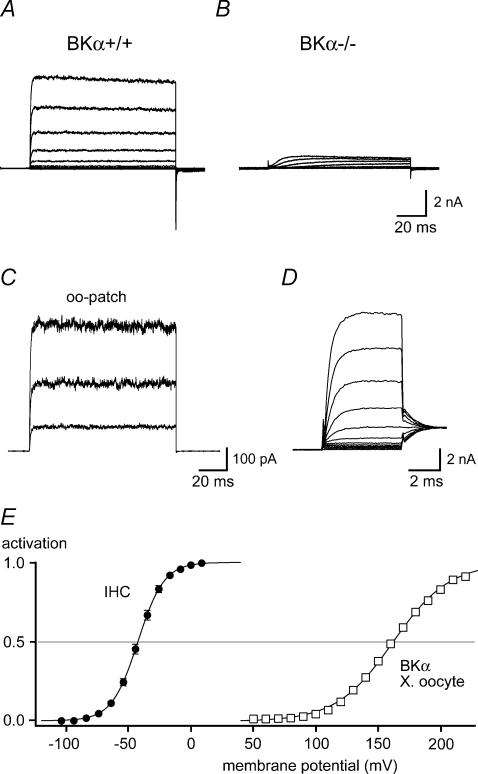
Figure 1. IK,f is mediated by BKCa channels.
A and B, outwardly rectifying K+ currents recorded in IHCs from a BKα knock-out mouse (BKα−/−; B) and a littermate control (BKα+/+; A). Currents were recorded in whole-cell mode in response to voltage steps to potentials between −104 and +6 mV (10 mV nominal increments) from a holding potential of −84 mV. Intracellular and extracellular solution contained 10 mm 4-AP and 1 μm XE991, respectively (see Methods). Note that the fast activating IK,f was absent in the BKα−/− IHC, where only a minor residual, slowly activating K+ current component was observed. C, BK currents recorded in an outside-out patch excised from a rat IHC. Current traces were evoked by voltage steps to +16, +56 and +96 mV, from a holding potential of −64 mV. Solutions were as in (A and B). Each trace is averaged from 10 individual presentations of the voltage step. D, fast activation of IK,f in a rat IHC, recorded as in A in response to 5-ms steps incremented by nominally 10 mV. Tail-current potential was −34 mV. Monoexponential fits to the current activation (not shown) yielded a time constant (τactivation) between 0.67 ms (at −44 mV) and 0.37 ms (at +6 mV). E, steady-state activation curve determined from tail currents measured in 16 rat IHCs as in C. Currents were normalized to the saturating tail-current amplitude and plotted versus the prepulse potential corrected for errors resulting from residual series resistance (see Methods); standard error of the voltage was smaller than the symbol size. The continuous line is a fit of a first-order Boltzmann function to the averaged data, yielding values for Vh and α of −42.4 mV and 10.4 mV, respectively. For comparison of the activation voltage ranges, activation curves obtained in excised patches from BKCa-expressing Xenopus oocytes at 0 [Ca2+]i are shown (□; mean data from seven patches; error bars are smaller than symbol size). The continuous line through the recombinant data (Xenopus oocyte) is a Boltzmann fit to the averaged data, yielding values for Vh and α of 161.9 mV and 22.6 mV, respectively.

