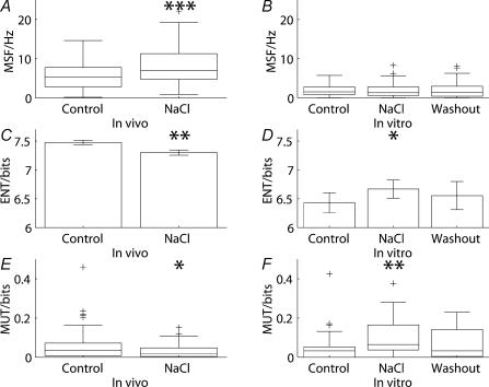
Figure 5. Responses to osmotic stimulation in vivo differed from those seen in vitro.
Before the hypertonic infusion of NaCl, 118 cells were recorded in vivo and 99 were recorded after the infusion whereas the number of cells tested in vitro was 30; no distinction was made between continuous and phasic cells. Where the distributions passed the normality test, means ±s.e.m. are represented; otherwise box and whiskers plots are shown. Osmotic stimulation in vivo significantly (z = 4.07, P < 0.001) increased (A) the mean spike frequency whereas the firing rate in vitro (B) was not significantly affected (z = −0.319, P = 0.750). The irregularity of activity, as measured by the log interval entropy, was decreased (C) in vivo (t = 2.92, P = 0.004) and increased (D) in vitro (t = 2.75, P = 0.010). Spike patterning, quantified using the mutual information between adjacent intervals, was decreased (E) in vivo (z = −2.40, P = 0.017) whereas the mutual information (F) in vitro was increased (z = −2.73, P = 0.006).

