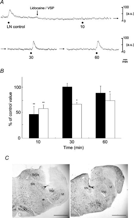
Figure 8. Effects produced by microinjection of lidocaine into the left (ipsilateral) trigeminal spinal nucleus (Vsp) and salivatory nuclei (SN) on the MBF increase elicited by left LN stimulation.
A, typical examples of the effects produced by microinjection of lidocaine (lignocaine; 4%) in a volume of 0.3 μl per site into the Vsp on the MBF increase elicited by LN stimulation (•), for 20 s with 20 V at 20 Hz using 2 ms pulses. B, the MBF increases evoked by LN stimulation 10, 30 and 60 min after microinjection of lidocaine into the Vsp (black bars, n = 6 in each group) and SN (white bars, n = 4 in each group) are expressed as percentages of the pretreatment response and given as means ± s.e.m. The statistical significance of the differences in this pretreatment response (control) was assessed by ANOVA followed by a post hoc test (Fisher's PLSD) (*P < 0.05, **P < 0.01, ***P < 0.001). C, photomicrographs of representative coronal sections stained with thionin through the medulla oblongata of the rat showing sites (arrowheads) at which microinjections of lidocaine were delivered to the Vsp (left panel) and SN (right panel). Scale bars represent 1 mm. FN, facial nucleus; IO, inferior olivary nucleus; SpVe, spinal vestibular nucleus; Vt, spinal trigeminal tract.

