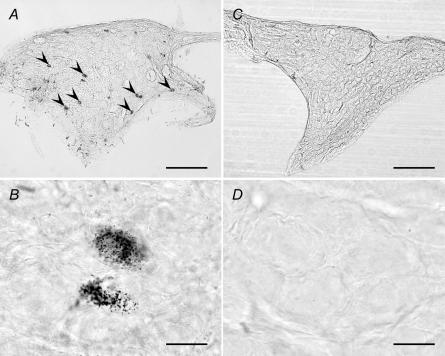
Figure 9. WGA–HRP-labelled neurones in the autonomic ganglions.
Photomicrographs showing wheat germ agglutinin–horseradish peroxidase (WGA–HRP)-labelled neurones after an injection of WGA–HRP into the masseter muscle in the rat. High- and low-power photomicrographs of the OG (A and B) and the pterygopalatine ganglion (PPG) (C and D) are shown. Arrowheads indicate the WGA–HRP-labelled cells. The left side of each photomicrograph is the rostral. Bars, 200 μm (A and C) and 20 μm (B and D).

