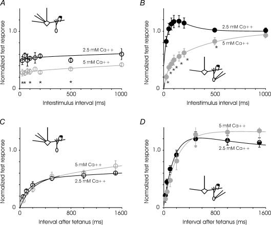
Figure 7. Calcium dependence of short-term plasticities.
A, paired-pulse depression of granule-to-mitral synapse in greater in high extracellular calcium (5 mm) compared to control conditions (2.5 mm). The time constant of recovery is not significantly different in the two cases. n = 6–14 cells for control, and 7 cells for high calcium. Data were fitted to the expression: 1 − 0.05e−t/100− 0.46e−t/6000 and 1 − 0.05e−t/180− 0.7e−t/6000 for low and high calcium, respectively. B, at higher calcium concentration, the mitral-to-granule synapse exhibits paired-pulse depression at all intervals tested. n = 6–12 cells for control and 7 cells for high calcium. Data were fitted to the expression: 1 − 1.2e−t/45+ 0.6e−t/150 and 1 − 0.4e−t/40− 0.6e−t/430 for low and high calcium, respectively. C, recovery from depression induced by fast trains was not substantially different at the granule-to-mtiral synapse in control (2.5 mm) and high (5 mm) calcium (n = 4 cells for each condition). Data were fitted to the expression: 1 − 0.4e−t/180− 0.5e−t/7000 and 1 − 0.4e−t/160− 0.6e−t/2000 for low and high calcium, respectively. D, recovery from depression at the mitral-to-granule synapse was also not affected significantly by increased extracellular calcium. (Control, n = 4 cells, high calcium, n = 5 cells.) Data were fitted to the expression: 1 − 1.45e−t/160+ 0.45e−t/1000 and 1 − 1.35e−t/200+ 0.35e−t/20000 for low and high calcium, respectively. Asterisks denote time points at which pair-wise comparisons within each panel reveals a difference at P < 0.05.

